There are many reasons you might choose to buy human anatomy atlas 3rd edition but it is not easy to find the best suitable human anatomy atlas 3rd edition for you. But don’t worry! We did some of the work for you already by researching a few models on the current market. Let’s check following article to find the best human anatomy atlas 3rd edition.
Best human anatomy atlas 3rd edition


Related posts:
Best human anatomy atlas 3rd edition reviews
1. Anatomy Flashcards
Description
Kaplan Medical's Anatomy Flashcards are designed to help you master the structures and systems of the human body. Whether you're a student, a health care practitioner, or just interested in learning about human anatomy, you can review on-the-go with 300 detailed, full-color cards.Essential Review
- 300 laminated flashcards with full-color, anatomically precise illustrations
- In-depth anatomical descriptions on the reverse side of each card
- Cards are organized by anatomical system with color-coded tabs for easy review
- 10 bonus coloring cards from Kaplan's top-selling Anatomy Coloring Book, so you can color, label, and learn each anatomical system for the ultimate in academic retention and recall
- We invented test prepKaplan (www.kaptest.com) has been helping students for almost 80 years. Our proven strategies have helped legions of students achieve their dreams.
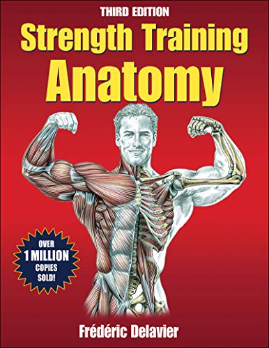
2. Strength Training Anatomy, 3rd Edition
Feature
HUMAN KINETICSDescription
With new exercises, additional stretches, and more of Frdric Delaviers signature illustrations, youll gain a whole new understanding of how muscles perform during strength exercises. This one-of-a-kind best-seller combines the visual detail of top anatomy texts with the best of strength training advice.
Many books explain what muscles are used during exercise, but no other resource brings the anatomy to life like Strength Training Anatomy. Over 600 full-color illustrations reveal the primary muscles worked along with all the relevant surrounding structures, including bones, ligaments, tendons, and connective tissue.
Like having an X-ray for each exercise, the anatomical depictions show both superficial and deep layers and detail how various setup positions affect muscle recruitment and emphasize underlying structures. New pages show common strength training injuries in a fascinating light and offer precautions to help you exercise safely.
Author and illustrator Frdric Delavier is the former editor in chief of the French publication PowerMag. He is a journalist for Le Monde du Muscle and a contributor to Mens Health Germany and several other strength training publications.
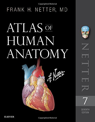
3. Atlas of Human Anatomy (Netter Basic Science)
Description
The only anatomy atlas illustrated by physicians, Atlas of Human Anatomy, 7th edition, brings you world-renowned, exquisitely clear views of the human body with a clinical perspective. In addition to the famous work of Dr. Frank Netter, youll also find nearly 100 paintings by Dr. Carlos A. G. Machado, one of todays foremost medical illustrators. Together, these two uniquely talented physician-artists highlight the most clinically relevant views of the human body. In addition, more than 50 carefully selected radiologic images help bridge illustrated anatomy to living anatomy as seen in everyday practice.
- Region-by-region coverage , including Muscle Table appendices at the end of each section.
- Large, clear illustrations with comprehensive labels not only of major structures, but also of those with important relationships.
- Tabular material in separate pages and additional supporting material as a part of the electronic companion so the printed page stays focused on the illustration.
Updates to the 7th Edition based on requests from students and practitioners alike:
- New Systems Overview section featuring brand-new, full-body views of surface anatomy, vessels, nerves, and lymphatics.
- More than 25 new illustrations by Dr. Machado, including the clinically important fascial columns of the neck, deep veins of the leg, hip bursae, and vasculature of the prostate; and difficult-to-visualize areas like the infratemporal fossa.
- New Clinical Tables at the end of each regional section that focus on structures with high clinical significance. These tables provide quick summaries, organized by body system, and indicate where to best view key structures in the illustrated plates.
- More than 50 new radiologic images some completely new views and others using newer imaging tools have been included based on their ability to assist readers in grasping key elements of gross anatomy.
- Updated terminology based on the international anatomic standard, Terminologia Anatomica, with common clinical eponyms included.
- Student Consult access includes a pincode to unlock the complete enhanced eBook of the Atlas through Student Consult. Every plate in the Atlasand over 100 Bonus Plates including illustrations from previous editionsare enhanced with an interactive label quiz option and supplemented with "Plate Pearls" that provide quick key points and supplemental tools for learning, reviewing, and assessing your knowledge of the major themes of each plate. Tools include 300 multiple choice questions, videos, 3D models, and links to related plates.
4. Atlas of Human Anatomy, Third Edition
Feature
Used Book in Good ConditionDescription
The ultimate anatomy atlas for medical study, clinical reference, and patient education, this updated masterpiece offers 534 of Netter's own accurate, clear and beautifully rendered illustrations along with eight Netter-style drawings rendered by Carlos A.G. Machado, MD. Netter's incomparable medical art and artistry reflects his personal belief in the power of the visual image to teach without overwhelming the student with dense, confusing text. "To clarify rather than intimidate" remains the distinctive and effective Netter approach and it's been working since the publication of the first edition in 1989.This masterwork has trained over 1,000,000 medical and health-science students
since its first release in 1989.
Updated with over 200 revised Netter illustrations, this new edition of the classic human anatomy atlas presents a total of 534 of Netter's own accurate, clear and precisely rendered illustrations along with 8 new Netter-style, surface anatomy drawings by Carlos A.G. Machado, MD.
This extraordinary new edition takes a major step forward to include surface anatomy and radiographic images to give a fuller, more integrated understanding of human anatomy. The index is expanded and improved, the references are updated, and a number of images are revised to reflect current knowledge. "To clarify rather than intimidate" continues to be the distinctive and effective Netter approach.
New Surface Anatomy Images - Each section begins with a surface anatomy plate to draw attention to the surface features that anticipate the underlying anatomy as well as highlight the value of careful observation in clinical medicine.
New Radiographic Images - For further investigation into anatomical detail
New and Revised Anatomical Images - Some plates were selected from Netter's Collection of Medical Illustrations 13-volume masterwork. Other images have been slightly revised and updated to reflect current knowledge.
Expanded and improved index and updated references
5. Netter's Atlas of Human Anatomy for CPT Coding, Third Edition
Description
This unique resource pairs the CPT code set and full descriptors with relevant anatomical illustrations created by famed medical illustrator and physician Frank Netter, MD, and others working in the Netter tradition. Each of seven chapters delves into a specific anatomical regionstarting with the head and ending with the lower extremitiesand opens with a brief introduction explaining the features of the region. The result is an illustrated explanation of how medical procedures relate to anatomy. This long-awaited third edition of Netters Atlas of Human Anatomy for CPT Coding provides readers a natural reference tool for reviewing clinical information and understanding the assignment of codes.
FEATURES AND BENEFITS
MORE THAN A DECADE OF CODE UPDATES! This third edition features CPT 2020 codes and descriptors from the Anesthesia, Surgery, Radiology, and Medicine sections of CPT Professional and integrates them with more than 500 anatomical illustrations from the Netter collection. The second edition featured the 2009 code set.
NEW ILLUSTRATIONS! New illustrations are featured in every chapter, with more than 100 new anatomical and procedural plates.
ADDITIONAL CODE SECTION INCLUDED! Category III codes are now included, giving visual context to the relationship between anatomy and emerging technology, services, procedures, and service paradigms.
Select CPT guidelines and instructional notes appear verbatim with the codes and illustrations while an appendix offers section opening guidelines for easy reference.
Nomenclature notes help explain related terminology.
A special symbol indicates when a procedural illustration exists for a code thereby allowing the reader to reference CPT Professional for additional information.
An appendix of abbreviations frequently found in CPT code descriptions and guidelines provide readers with information that can aid them in understanding documentation used to support code assignment.
NEW EPONYMS APPENDIX! A procedural eponyms appendix gives not only a list of common eponyms found within the code set but a brief explanation of each procedure.
NEW PROCEDURAL ILLUSTRATIONS APPENDIX! The inclusion of select procedural illustrations found in
CPT Professional helps readers make the visual connects between anatomy and procedure.
6. Photographic Atlas for Anatomy & Physiology, A
Description
For 2-semester A&P lab course and 1-semester human anatomy lab course
A Photographic Atlas for Anatomy & Physiology is a new visual lab study tool that helps students learn and identify key anatomical structures. Featuring photos from Practice Anatomy Lab 3.1 and other sources, the Atlas includes over 250 cadaver dissection photos, histology photomicrographs, and cat dissection photos plus over 50 photos of anatomical models from leading manufacturers such as 3B Scientific, SOMSO, and Denoyer-Geppert Science Company. Two-page spreads with cadaver and anatomical model photos side-by-side help students to better learn and identify structures. The Atlas is composed of 13 chapters, organized by body system, and includes a final chapter with cat dissection photos. In each chapter, students will first explore gross anatomy, as seen on cadavers and anatomical models, and then conclude with relevant histological images.
7. A Brief Atlas of the Human Body
Feature
spiral boundDescription
This full-color atlas includes 107 bone and 47 soft-tissue photographs with easy-to-read labels. This new edition of the atlas contains a brand new comprehensive histology photomicrograph section featuring over 50 slides of basic tissue and organ systems. Featuring photos taken by renowned biomedical photographer Ralph Hutchings, this high-quality photographic atlas makes an excellent resource for the classroom and laboratory, and is referenced in appropriate figure legends throughout the text.8. Atlas of Anatomy
Description
For the student just starting on their medical journey or for a neurosurgeon looking to replace an outdated atlas in his or her library, this is a wonderful option. -- YNC Newsletter (Young Neurosurgeons News)
Highly recommended to students and surgeons....Of very high technical quality -- Pediatric Endocrinology Reviews
With unmatched accuracy, quality, and clarity, the Atlas of Anatomy is now fully revised and updated.
Atlas of Anatomy, Third Edition, is the highest quality anatomy atlas available today. With over 1,900 exquisitely detailed and accurate illustrations, the Atlas helps you master the details of human anatomy.
Key Features:
- NEW! Sectional and Radiographic Anatomy chapter for each body region
- NEW! Radiologic images help you connect the anatomy lab to clinical knowledge and practice
- NEW! Pelvis and Perineum section enhanced and improved making it easier to comprehend one of the most complex anatomic regions
- NEW! Section on Brain and Nervous System focuses on gross anatomy of the peripheral and autonomic nervous systems as well as the brain and central nervous system
- Also included in this new edition:
- More than 170 tables summarize key details making them easier to reference and retain
- Muscle Fact spreads provide essential information, including origin, insertion, innervation, and action
- An innovative, user-friendly format: every topic covered in two side by side pages
- Access to WinkingSkull.com PLUS, with all images from the book for labels-on and labels-off review and timed self-tests for exam preparation
What students say about the Atlas of Anatomy:
"Thieme is the best anatomy atlas by far, hands down. Clearer pictures, more pictures, more realistic pictures, structures broken up in ways that make sense and shown from every angle...includes clinical correlations...That's about all there is to it. Just buy it. Thank you Thieme!"
"...this book surpasses them all. It's the artwork. The artist has found the perfect balance of detail and clarity. Some of these illustrations have to be seen to be believed.... The pearls of clinical information are very good and these add significance to the information and make it easier to remember."
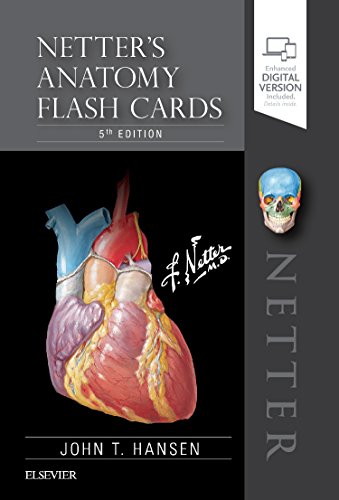
9. Netter's Anatomy Flash Cards (Netter Basic Science)
Description
Learn the essential anatomy you need to know quickly and easily! Each flash card in this full-color deck features high-quality Netter art (and several new paintings by Dr. Carlos Machado), numbered labels (with hidden answers), and concise comments and clinical notes for the most commonly tested anatomy terms and concepts. Focusing on clinically relevant anatomy, this easy-to-use, portable study tool helps you learn anatomical structures with confidence!
- Pre-punched holes make it easy to carry selected groups of cards with you.
- A perfect study aid and complement to Netters Clinical Anatomy, 4rd Edition concise textbook and Netters Atlas of Human Anatomy, 7th Edition.
- New card design makes it easy to sort cards by Region (primary color-coded organization) or System (icons).
- Student Consult eBook version included with purchase. This enhanced eBook experience allows you to study the cards on your phone, tablet, or computer and includes over 400 multiple-choice questions. Quiz yourself on structure names as well as their anatomical and clinical significance.
10. Human Sectional Anatomy: Pocket atlas of body sections, CT and MRI images, Fourth edition
Description
First published in 1991, Human Sectional Anatomy set new standards for the quality of cadaver sections and accompanying radiological images. Now in its fourth edition, this unsurpassed quality remains and is further enhanced by the addition ofnew material.
The superb full-colour cadaver sections are compared with CT and MRI images, with accompanying, labelled, line diagrams. Many of the radiological images have been replaced with new examples for this latest edition, captured using the most up-to date imaging technologies to ensure excellent visualization of the anatomy. The photographic material is enhanced by useful notes with details of important anatomical and radiological features.
Beautifully presented in a convenient and portable format, the fourth edition of this popular pocket atlas continues to be an essential textbook for medical and allied health students and those taking postgraduate qualifications in radiology, surgery and medicine, and an invaluable ready-reference for all practising anatomists, radiologists, radiographers, surgeons and medics.