Finding your suitable atlas of human anatomy is not easy. You may need consider between hundred or thousand products from many store. In this article, we make a short list of the best atlas of human anatomy including detail information and customer reviews. Let’s find out which is your favorite one.
Best atlas of human anatomy


Related posts:
Best atlas of human anatomy reviews
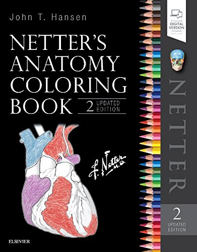
1. Netter's Anatomy Coloring Book Updated Edition (Netter Basic Science)
Description
Learn and master anatomy with ease, while having fun, through the unique approach of Netter's Anatomy Coloring Book, 2nd Edition. You can trace arteries, veins, and nerves through their courses and bifurcations...reinforce your understanding of muscle origins and insertions from multiple views and dissection layers...and develop a better understanding of the integration of individual organs in the workings of each body system throughout the human form. Whether you are taking an anatomy course or just curious about how the body works, let the art of Netter guide you!
Key Features
- Netter's Anatomy Coloring Book is a perfect companion to the Atlas of Human Anatomy by Frank H. Netter, MD as well as Netter's Anatomy Flash Cards and Netter's Clinical Anatomy textbook.
- Understand the correlation between structures. Outlines of Netter anatomical illustrations in multiple views, magnifications, and dissection layers, accompanied by high-yield information reinforce visual recognition and provide context.
- Master challenging structures through illustrations small enough for quick coloring, but large enough to provide you with important details.
- Facilitate learning by following tips for coloring key structures and quizzing yourself with end-of-section review questions.
- Quickly review key concepts with accompanying tables that review muscle attachments, innervation, and actions.
- Understand the role of anatomy in medicine through Clinical Notes which highlight examples.
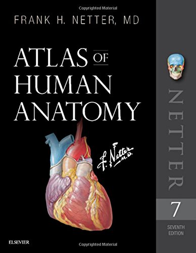
2. Atlas of Human Anatomy (Netter Basic Science)
Description
The only anatomy atlas illustrated by physicians, Atlas of Human Anatomy, 7th edition, brings you world-renowned, exquisitely clear views of the human body with a clinical perspective. In addition to the famous work of Dr. Frank Netter, youll also find nearly 100 paintings by Dr. Carlos A. G. Machado, one of todays foremost medical illustrators. Together, these two uniquely talented physician-artists highlight the most clinically relevant views of the human body. In addition, more than 50 carefully selected radiologic images help bridge illustrated anatomy to living anatomy as seen in everyday practice.
- Region-by-region coverage , including Muscle Table appendices at the end of each section.
- Large, clear illustrations with comprehensive labels not only of major structures, but also of those with important relationships.
- Tabular material in separate pages and additional supporting material as a part of the electronic companion so the printed page stays focused on the illustration.
Updates to the 7th Edition based on requests from students and practitioners alike:
- New Systems Overview section featuring brand-new, full-body views of surface anatomy, vessels, nerves, and lymphatics.
- More than 25 new illustrations by Dr. Machado, including the clinically important fascial columns of the neck, deep veins of the leg, hip bursae, and vasculature of the prostate; and difficult-to-visualize areas like the infratemporal fossa.
- New Clinical Tables at the end of each regional section that focus on structures with high clinical significance. These tables provide quick summaries, organized by body system, and indicate where to best view key structures in the illustrated plates.
- More than 50 new radiologic images some completely new views and others using newer imaging tools have been included based on their ability to assist readers in grasping key elements of gross anatomy.
- Updated terminology based on the international anatomic standard, Terminologia Anatomica, with common clinical eponyms included.
- Student Consult access includes a pincode to unlock the complete enhanced eBook of the Atlas through Student Consult. Every plate in the Atlasand over 100 Bonus Plates including illustrations from previous editionsare enhanced with an interactive label quiz option and supplemented with "Plate Pearls" that provide quick key points and supplemental tools for learning, reviewing, and assessing your knowledge of the major themes of each plate. Tools include 300 multiple choice questions, videos, 3D models, and links to related plates.
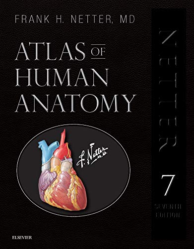
3. Atlas of Human Anatomy, Professional Edition: including NetterReference.com Access with Full Downloadable Image Bank (Netter Basic Science)
Description
The only anatomy atlas illustrated by physicians, Atlas of Human Anatomy, 7th edition, brings you world-renowned, exquisitely clear views of the human body with a clinical perspective. In addition to the famous work of Dr. Frank Netter, youll also find nearly 100 paintings by Dr. Carlos A. G. Machado, one of todays foremost medical illustrators. Together, these two uniquely talented physician-artists highlight the most clinically relevant views of the human body. In addition, more than 50 carefully selected radiologic images help bridge illustrated anatomy to living anatomy as seen in everyday practice.
- Region-by-region coverage , including Muscle Table appendices at the end of each section.
- Large, clear illustrations with comprehensive labels not only of major structures, but also of those with important relationships.
- Tabular material in separate pages and additional supporting material as a part of the electronic companion so the printed page stays focused on the illustration.
Updates to the 7th Edition based on reader requests:
- Full downloadable image bank, now with on/off functionality for individual labels, available at NetterReference.com.
- New Systems Overview section featuring brand-new, full-body views of surface anatomy, vessels, nerves, and lymphatics.
- More than 25 new illustrations by Dr. Machado, including the clinically important fascial columns of the neck, deep veins of the leg, hip bursae, and vasculature of the prostate; and difficult-to-visualize areas like the infratemporal fossa.
- New Clinical Tables at the end of each regional section that focus on structures with high clinical significance. These tables provide quick summaries, organized by body system, and indicate where to best view key structures in the illustrated plates.
- More than 50 new radiologic images some completely new views and others using newer imaging tools have been included based on their ability to assist readers in grasping key elements of gross anatomy.
- Updated terminology based on the international anatomic standard, Terminologia Anatomica, with common clinical eponyms included.
- Purchase of this Professional Edition allows access to the full downloadable image bank of the current Atlas as well as additional plates from previous editions and other bonus content at NetterReference.com. [*Your Registered User License allows for the creation of presentations for your individual, personal use which you can present in small group settings of 10 or fewer people. It also permits registered student users to include images in posters at scientific conferences as long as proper citation is included. Complete Registered User License as well as contact information for Institutional sales can be found at www.NetterReference.com.] Access will terminate 5 years from publication or upon publication of the next edition of this title.
4. Jean Marc Bourgery. Atlas of Human Anatomy and Surgery (Bibliotheca Universalis)
Feature
Bourgery Atlas of Human Anatomy and SurgeryDescription
We owe a great debt to Jean Baptiste Marc Bourgery (17971849) for his Atlas of Anatomy, which was not only a massive event in medical history, but also remains one of the most comprehensive and beautifully illustrated anatomical treatises ever published.
Bourgery began work on his magnificent atlas in 1830 in cooperation with illustrator Nicolas Henri Jacob (17821871), a student of the French painter Jacques Louis David. The first volumes were published the following year, but completion of the treatise required nearly two decades of dedication; Bourgery lived just long enough to finish his labor of love, but the last of the treatises eight volumes was not published in its entirety until five years after his death.
The eight volumes of Bourgerys treatise cover descriptive anatomy, surgical anatomy and techniques (exploring in detail nearly all the major operations that were performed during the first half of the 19th century), general anatomy and embryology, and microscopic anatomy. Jacobs spectacular hand-colored lithographs are remarkable for their clarity, color, and aesthetic appeal, reflecting a combination of direct laboratory observation and illustrative research. Unsurpassed to this day, the images offer exceptional anatomical insight, not only for those in the medical field but also for artists, students, and anyone interested in the workings and wonder of the human body.
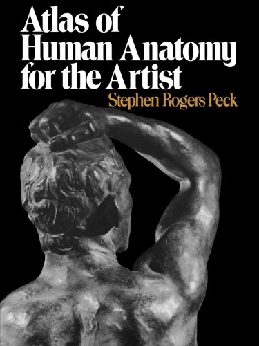
5. Atlas of Human Anatomy for the Artist
Feature
Atlas of Human Anatomy for the ArtistDescription
Stephen Rogers Peck's Atlas of Human Anatomy for the Artist remains unsurpassed as a manual for students. It includes sections on bones, muscles, surface anatomy, proportion, equilibrium, and locomotion. Other unique features are sections on the types of human physique, anatomy from birth to old age, an orientation on racial anatomy, and an analysis of facial expressions. The wealth of information offered by the Atlas ensures its place as a classic for the study of the human form.6. Anatomy: A Photographic Atlas (Color Atlas of Anatomy a Photographic Study of the Human Body)
Feature
Anatomy A Photographic AtlasDescription
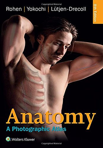
Prepare for the dissection lab and operating room with Anatomy: A Photographic Atlas, 8e. Featuring outstanding full-color photographs of actual cadaver dissections with accompanying schematic drawings and diagnostic images, this proven text depicts anatomic structures more realistically than illustrations in traditional atlases. Chapters are organized by region in the order of a typical dissection with each chapter presenting topographical anatomical structures in a systemic manner.
- Authentic photographic reproduction of colors, structures, and spatial dimensions as seen in the dissection lab and on the operating table help you develop an understanding of the anatomy of the human body.
- Functional connections between single organs, the surrounding tissue, and organ systems are clarified to prepare you for the dissection lab and practical exams.
- Clinical cases and over 1,200 images enhance your understanding.
- Dissections illustrate the topographical anatomy in layers "from the outside in" to better prepare you for the lab and operating room.
7. Atlas of Human Anatomy
Description
This new manual takes a systemic approach with each chapter focusing on one body system. The order of chapters will follow the traditional order found in anatomy or anatomy and physiology courses. The photos will include skeletal images, photomicrographs of histology and cadaver dissections. This atlas includes full-color photographs of actual cadaver dissections instead of idealized illustrations, to accurately and realistically represent anatomical structures. The goal is to produce an atlas that is a strong teaching and learning publication and not just a catalog of photographs.8. The Color Atlas of Human Anatomy
Feature
Sterling Publishing NYDescription
9. Atlas of Human Anatomy, 4th Edition (Netter Basic Science)
Feature
Used Book in Good ConditionDescription
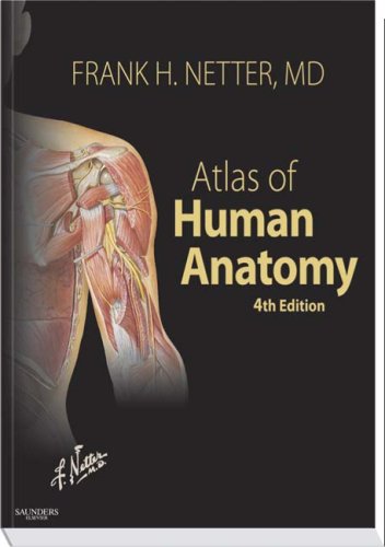
Netter's Atlas of Human Anatomy is the most loved and best selling anatomy atlas in the English language. In over 540 beautifully colored and easily understood illustrations, it teaches the complete human body with unsurpassed clarity and accuracy. This new edition features 45 revised, 290 relabeled and 17 wholly new plates, drawn fully in the tradition of Frank Netter, and includes more imaging and clinical images than ever before. Six Consulting Editors have worked together to ensure the new edition's accuracy and usefulness in the lecture theatre, classroom and dissection lab. Ninety plates from the book as well as a powerful and varied bank of ancillary material, unique to this atlas, are available online through www.netteranatomy.com.- Includes uniquely informative drawings that allow you - and have allowed generations of students - to learn structures with confidence.
- Associates normal anatomy with an application of that knowledge in a clinical setting.
- Offers a strong selection of imaging to show you what is happening three dimensionally in the human body, the way you see it in practice.
At www.netteranatomy.com, you'll access...
Over 90 of the most important anatomy illustrations from the book.
Interactive Anatomy Dissection Modules.
Radiographs, CT scans, MRIs, and angiograms,
with "labels on/off" buttons for self testing.
QuickTime movies of stacked, transverse, and sectional images
from the Visible Human Project (VHP).
Review Center with "Identification Spot Tests" and USMLE-style
multiple-choice questions.
Integration links to other STUDENT CONSULT titles.
and more!
10. Atlas of Human Anatomy: with Student Consult Access (Netter Basic Science)
Feature
Includes Online Student ConsultDescription
Atlas of Human Anatomy uses Frank H. Netter, MD's detailed illustrations to demystify this often intimidating subject, providing a coherent, lasting visual vocabulary for understanding anatomy and how it applies to medicine. This fifth edition features a stronger clinical focus-with new diagnostic imaging examples--making it easier to correlate anatomy with practice. Student Consult online access includes supplementary learning resources, from additional illustrations to an anatomy dissection guide and more. Netter. It's how you know.
- See anatomy from a clinical perspective with hundreds of exquisite, hand-painted illustrations created by, and in the tradition of, pre-eminent medical illustrator Frank H. Netter, MD.
- Join the global community of healthcare professionals who've mastered anatomy the Netter way!
- Expand your study at Student Consult online, where you'll find a suite of learning aids including selected Netter illustrations, additional clinically focused illustrations and radiologic images, videos from Netter's 3D Interactive Anatomy, dissection modules, an anatomy dissection guide, multiple-choice review questions, "drag-and-drop" exercises, clinical pearls, clinical cases, survival guides, surgical procedures, and more.
- Correlate anatomy with practice through an increased clinical focus, many new diagnostic imaging examples, and bonus clinical illustrations and guides online.


